Research Highlights
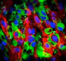
When our drugs don't work.
Eribulin was developed as a potent anticancer agent, but it fails in many patients for unknown reasons. In a recent study, CSB researchers used microscopic imaging in tumors to show that resistance is primarily due to MDR1-mediated drug efflux. It was discovered that a new nano-encapsulated MDR1 inhibitor was able to restore drug efficacy. These studies show that in vivo imaging is a powerful strategy for elucidating mechanisms of drug resistance in heterogeneous tumors and for evaluating strategies to overcome this resistance.
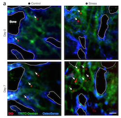
Stem cells get stressed out too!
For decades, doctors knew that chronic stress is bad for you. The study by Heidt and Sager found that psychosocial stress activates bone marrow stem cells, which in turn triggers overproduction of inflammatory leukocytes, including neutrophils and monocytes. These leukocytes are more numerous in blood and accumulate in atherosclerotic lesions, putting the individual at higher risk for myocardial infarction and stroke.
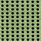
Tiny holes enable big measurements
A new technology developed at CSB allows profiling of small subcellular structures such as exosomes. The technology uses tiny gold grids studded with nanoholes in array format. Each of the holes has been modified with different antibodies. Biomarkers interacting with these tiny nano holes change the light properties (nano-plasmons), an effect which can optically detected. This technology (”nPLEX”) will allow high-throughput analysis of a number of clinically important biomarkers.
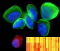
More (proteomic) bang-for-your-buck
Extracting maximal information from minimal, easily acquired samples is the holy grail for patient monitoring in clinical trials. A new technology developed at the CSB, holds promise for revolutionizing clinical monitoring by allowing vast amounts of proteomic information to be obtained from minute samples.
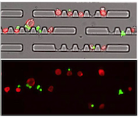
Repurposing often discarded ascites
Buildup of cancer-containing fluid in the abdomen (ascites) is a common occurrence suffered by many patients with advanced malignancies. Researchers have developed and tested a novel microfluidic chip, reported in PNAS, to selectively detect ascites tumor cells and enable further testing. This approach seeks to repurpose ascites for serial pharmacodynamic readouts.
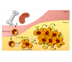
Proliferation rules
According to a recent study by CSB researchers, the primary driver of atherosclerotic plaque growth is local proliferation of plaque-resident inflammatory cells. This finding not only challenges previous assumptions that plaque growth is the exclusive result of cell recruitment from the blood, but now raises hope for the future development of targeted atherosclerotic treatment. Findings are reported in Nature Medicine.

Cracking bacterial code for quick diagnosis
Researchers have developed a quick and sensitive method for identifying and characterizing infectious bacteria in patient samples. The genetic test uses specially designed DNA probes that seek out and magnetically label regions of bacterial RNA. Reported in Nature Nanotechnology, this diagnostic technique is powerful enough to detect and differentiate single bacteria in under 2 hours.
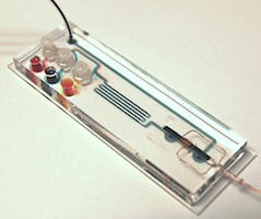
Faster cheaper TB diagnosis
A nanotechnology inspired genetic test has been developed to detect tuberculosis (TB) directly in sputum. The method not only distinguishes different forms of TB but also sheds light on drug resistant strains. The innovative device, described in Nature Communications, is also capable of comprehensive diagnosis in two hours and is sensitive enough to detect single bacteria.
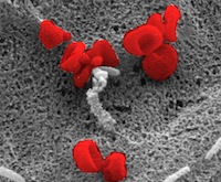
The magic of “cell dust”
A novel diagnostic platform has been shown capable of detecting minuscule particles shed by cells, known as microvesicles, in a drop of blood. In a groundbreaking study, published in Nature Medicine, we demonstrate that by using nanotechnology together with nuclear magnetic resonance (NMR), microvesicles shed by brain cancer cells can be reliably detected in human blood samples.
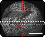
Imaging the beating heart in the living body
Imaging individual cells in a live beating heart has been nearly impossible. Yet, this would offer unprecedented opportunities to better understand heart diseases. A new imaging method - reported in the October issue of Nature Communications - and developed by researchers at the Center for System Biology now allows single cell resolution imaging in cardiovascular research.

The magic of “cell dust”
A novel diagnostic platform has been shown capable of detecting minuscule particles shed by cells, known as microvesicles, in a drop of blood. In a groundbreaking study, published in Nature Medicine, investigators at the Center for Systems Biology (CSB) demonstrate that by using nanotechnology together with nuclear magnetic resonance (NMR), microvesicles shed by brain cancer cells can be reliably detected in human blood samples.
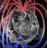
Finding a needle in a haystack
Researchers have developed a novel microchip that can rapidly scan through the enormous number of cells in a blood sample to find very rare circulating tumor cells. Using a combination of microelectronics, microfluidics, and nanotechnology, this new system, described in the July issue of Science Translational Medicine, now has the potential for rapid and accurate on-the-spot cancer diagnosis and treatment monitoring.
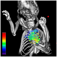
Not just a ‘plumbing’ disease
New research at the Center for Systems Biology (CSB) has shown that heart attacks are not just a “plumbing” problem in the arteries but a ‘whole system’ condition, resulting in widespread inflammation and a predisposition for a secondary attack.
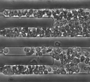
Sickle symptoms linked to cell flow
The genetic abnormality that causes blood cells to deform and clog vessels in sickle cell disease results in wide symptom variability across patients. Until now, there has been no way to distinguish patients with severe from patients with mild disease. A novel technique, however, has shown promise in differentiating patients based on the rate at which their blood stops flowing under low oxygen.
A new cell discovered
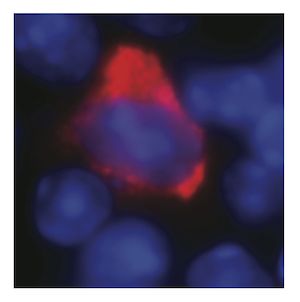
Sepsis is a life-threatening condition characterized by whole-body inflammation to overwhelming infection. A newly discovered cell type known as “innate response activator” has been found to protect against sepsis. This discovery will likely provide insight into the development of sepsis and potentially lead to new avenues for therapeutic intervention.
Novel therapy turns off navigation system in disease-promoting cells
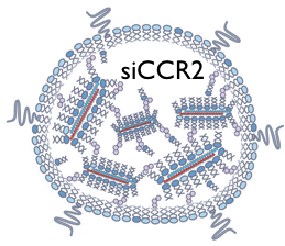
By manipulating the molecular navigation system used by inflammatory immune cells to reach sites of tissue damage, researchers at the Center for Systems Biology may have struck upon an effective novel anti-inflammatory treatment. Given that inflammation is an exacerbator of almost all major diseases, this therapy could potentially have wide-spread benefit.
Probing the heart for infection
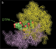
A new imaging probe developed by researchers at the Center for Systems Biology can detect acute endocarditis, a rapidly progressing infection of the heart valves. This imaging agent binds tightly to a product released by the most deadly cause of the infection-Staphylococcus aureus-rendering it visible by both optical and PET imaging. Ultimately, the agent could be used to rapidly diagnose, and thus treat, this potentially fatal condition.

The spleen's newly discovered function
While the spleen has long held a reputation for redundancy, recent research has now shown that quite the opposite is true. The spleen, in fact, appears to play an important role in the repair of tissue. Mikael Pittet, one of the four lead investigators responsible for this finding, discusses this work and its possible implications.
Glow-in-the-dark plaques
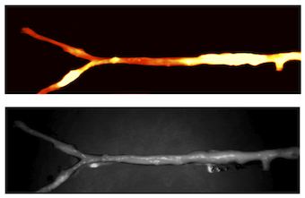
Finding new uses for already FDA approved drugs is a speedy way of translating new biological discoveries into patient benefit. Using a novel catheter-based imaging system, the fluorescent dye Indocyanine green has been shown capable of highlighting the culprits responsible for strokes and heart attacks, namely inflamed arterial plaques.
When small meets speedy...
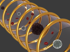
In today's fast paced world, waiting for anything is often frustrating and stressful. But few delays can be worse than that following a diagnostic blood test or biopsy, where results can take days to come back. Recently, a new portable device, known as DMR has been shown capable of on the spot cancer diagnosis.