
Bioengineering Can Do It! The cover image symbolizes the process of translating bioengineering research from the laboratory to the clinic. In this Special Issue focused on translational bioengineering, an Editorial (Weissleder) discusses impediments and ideas to improve commercialization of biotechnology. Three Review articles discuss translational challenges and advances in medical wearables (Xu et al.), musculoskeletal tissue regeneration (Khodabukus et al.), and diagnostics and drug delivery for low-resource environments (Euliano et al.). A Viewpoint (Bliley et al.) highlights innovations in three-dimensional (3D) bioprinting and forecasts key milestones for clinical translation. Three Research Articles complete the Special Issue, presenting 3D-printed in-line ventilators for acute lung injury (Pritchard et al.), a microparticle therapy for arthrofibrosis (Kirsch et al.), and engineered mini-bones for studying cancer metastasis (Grigoryan et al.). This Special Issue showcases the pitfalls, progress, and potential of translational bioengineering. Credit: Jason Solo/The Jacky Winter Group

Pancreatic cancer spatial transcriptomics
The lethality and treatment-refractory nature of pancreatic cancer are largely mediated by collaborative interactions between cancer cells and other cell types in the tumor microenvironment, including cancer-associated fibroblasts and immune cells. By constructing a high-resolution molecular landscape of the multicellular subtypes and spatial communities that compose pancreatic cancer and the dynamic remodeling associated with cytotoxic selection pressure, additional therapeutic vulnerabilities are identified to augment precision oncology efforts in pancreatic cancer.
Extracellular vesicles (EVs) carry information inherited from parental cells, having significant potential for disease diagnosis. In blood, however, EVs are outnumbered >104-fold by low density lipoproteins (LDLs), yet similar in size and density. These fundamental disadvantages often cause LDL spillover into EV isolates, thus confounding assay results. We hypothesized that EVs can be further separated from LDLs based on electric charge: EVs and LDLs have different lipid composition, which can lead to differential surface charge densities. To test this hypothesis, we modeled and quantified the surface charge of EVs and LDLs, and used the information to optimally separate EVs from LDLs via ion-exchange chromatography.

EPOCHal Test. Current tests able to detect and quantify tetrahydrocannabinol (THC), the psychoactive ingredient of marijuana (pictured), in bodily fluids are routinely used for drug use prevention and treatment. These tests suffer from low sensitivity and take too long. Yu et al. have developed a sensitive test, called EPOCH (express probe for on-site cannabis inhalation), that allows THC quantification in saliva on-site in minutes. EPOCH was tested in marijuana users and provided reproducible results with a sensitivity compatible with regulatory guidelines. EPOCH represents a fast, effective, and noninvasive method to monitor THC in bodily fluids.
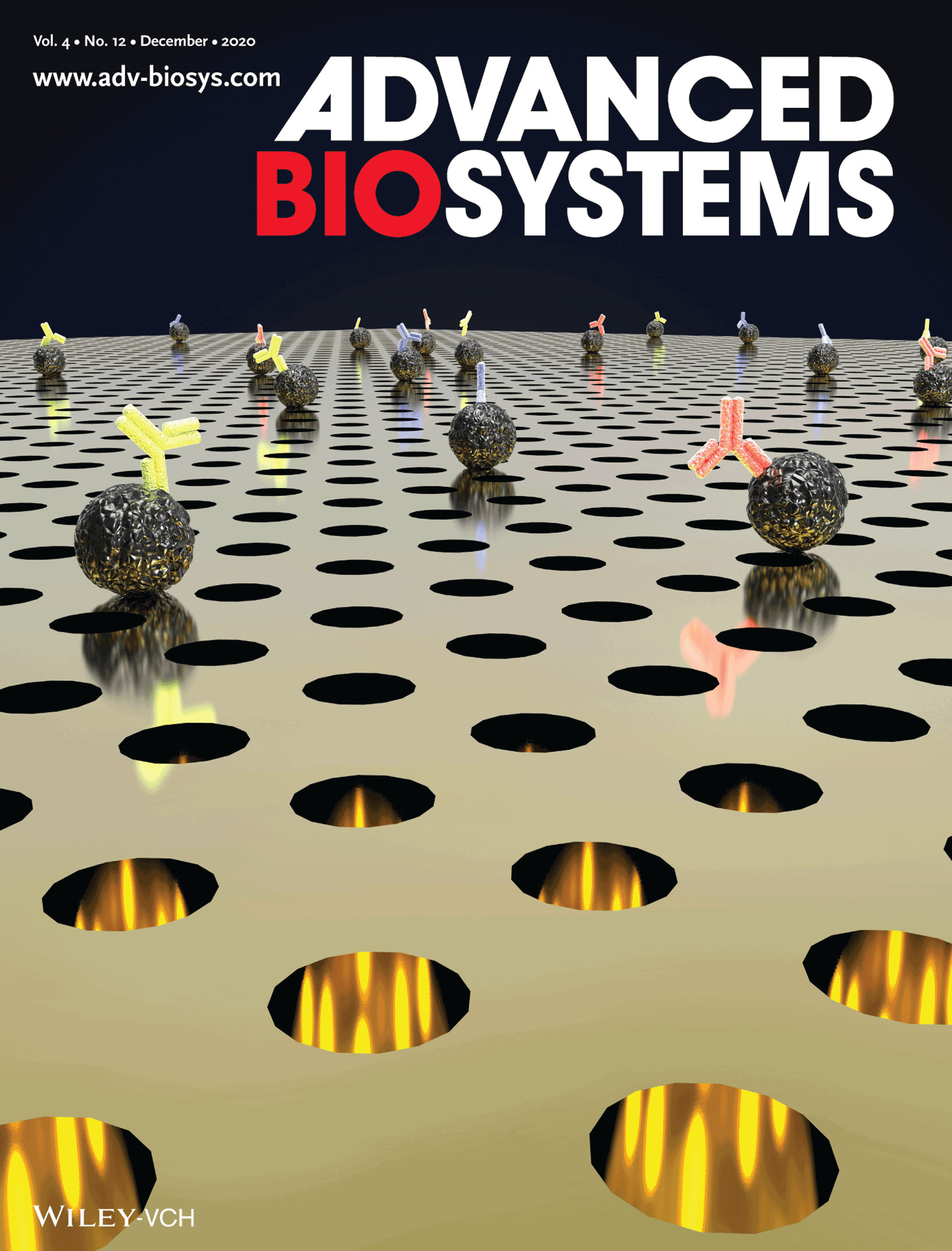
In article Plasmon‐Enhanced Biosensing: Plasmon‐Enhanced Biosensing for Multiplexed Profiling of Extracellular Vesicles Hyungsoon Im and co‐workers label extracellular vesicles, which are nanoscale phospholipid vesicles secreted by cells, with fluorescently conjugated antibodies. The fluorescence signals are amplified by periodic gold nanohole structures based on surface plasmon resonance. Multi‐fluorescence imaging enables multiplexed analyses of proteins with improved sensitivities.

Radiotheranostics, injectable radiopharmaceuticals with antitumour effects, have seen rapid development over the past decade. Although some formulations are already approved for human use, more radiopharmaceuticals will enter clinical practice in the next 5 years, potentially introducing new therapeutic choices for patients. Despite these advances, several challenges remain, including logistics, supply chain, regulatory issues, and education and training. By highlighting active developments in the field, this Review aims to alert practitioners to the value of radiotheranostics and to outline a roadmap for future development. Multidisciplinary approaches in clinical trial design and therapeutic administration will become essential to the continued progress of this evolving therapeutic approach.
In this article, Sung‐Gyu Park, Hyungsoon Im, Dong‐Ho Kim, and co‐workers develop a direct formation and selective self‐assembly of spherical plasmonic nanoparticles on slippery 3D Au nanopillars through a simple vacuum deposition process by enhancing the surface diffusion of adsorbed plasmonic atoms. The 3D plasmonic chips show very high performance in both surface‐enhanced Raman spectroscopy and plasmon‐enhanced fluorescence for avian influenza detection.
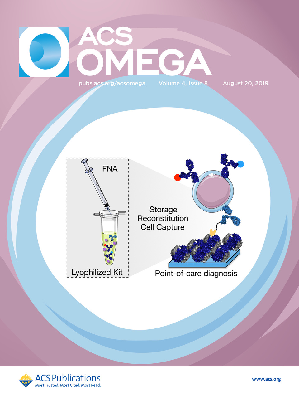
This image shows a simple method for the immobilization of cells on glass surfaces. The methodology significantly improves the ability of the neutravidin-coated glass slides to withstand lyophilization and storage.
For more information, see Marquard AN, Carlson JCT, Weissleder R, pp 11515−11521.

Luminal alkaline pH increases the length of V-ATPase-labeled microplicae in epididymal proton-secreting clear cells. Cauda epididymis was luminally perfused in vivo with a solution adjusted to the alkaline pH of 7.8. 3D reconstruction by Airyscan microscopy shows numerous long V-ATPase-labeled microplicae (green) in this “activated” clear cell. Double-labeling for the endocytic marker clathrin (red) was performed to identify the apical border of the clear cell and adjacent principal cells. Nuclei are labeled in blue with the DNA marker DAPI. See Battistone et al. pp 1957–1973.

Immunocytochemistry shows the distribution of the proto-oncogene tyrosine-protein kinase Src (green), and the vasopressin-sensitive water channel (AQP2, red) in rat inner medullary principal cells. Src inhibition induces an AQP2 membrane accumulation that requires the phosphorylation of serine 269 but not serine 256, the well-known master key phosphorylation serine residue on the AQP2 c-terminus. Our study shows an independent pathway that can target different AQP2 residues, and help to design new strategies to induce or sustain AQP2 membrane expression when VP signaling is defective. See Cheung et al. pp 1627–1642.

‘Heart of a snowman’, inspired by Swirski FK and Nahrendorf M.
The past few decades have generated growing recognition that the immune system makes an important contribution to cardiac development, composition and function. Immune cells infiltrate the heart at gestation and remain in the myocardium, where they participate in essential housekeeping functions throughout life. After myocardial infarction or in response to infection, large numbers of immune cells are recruited to the heart to remove dying tissue, scavenge pathogens and promote healing. Under some circumstances, immune cells can cause irreversible damage, contributing to heart failure. This Review focuses on the role of the immune system in the heart under both homeostatic and perturbed conditions.

Lights, camera, action.
Pittet et al. examine the fundamentals of using intravital microscopic imaging to study single cells and highlight how this technology has allowed us to directly visualize the movement and activities of immune cells in various tissues. They also provide a comprehensive overview of mouse reporter lines that are currently available to monitor immune cell dynamics using intravital imaging. Featured on the cover is a hand-drawn sketch of a microscopy room used for intravital imaging.

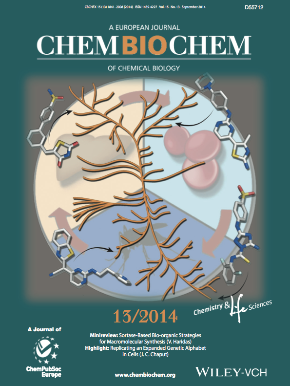
A ghost in the mechanism: tetrazine/trans-cyclooctene click-to-release chemistry has driven recent developments in bioorthogonal bond cleavage, but has proven difficult to optimize. Unraveling the post-click reaction network, the “release cube”, revealed a previously overlooked tricyclic dead-end isomer and enabled strategies to block its formation to achieve complete and ultrafast release. See the article by Carlson et al

Hulsmans et al. report that cardiac macrophages expand in left ventricular diastolic dysfunction, a hallmark of heart failure with preserved ejection fraction (HFpEF) and cardiac aging. In HFpEF, macrophages shift toward a profibrotic subset that promotes ventricular stiffness. The cover illustrates Mac3+ macrophages in the left ventricle of a mouse with hypertension-induced diastolic dysfunction.

Antibodies against immune checkpoint proteins such as PD-1 keep antitumor T cells from becoming exhausted and can greatly improve the survival of cancer patients, but even this therapy is not always effective, and a paper by Arlauckas et al. demonstrates one reason why. Using intravital imaging to track individual cells and antibodies, the authors observed macrophages removing anti–PD-1 antibodies from T cells, thus inactivating them. This image shows an example of this interaction, with a macrophage (red) directly contacting T cells (blue) and removing the anti–PD-1 antibody (yellow).

Immunofluorescence microscopy of liver from Rosa26CAG-tdTomato Alb-Cre+ reporter mice demonstrates Cre-mediated recombination in hepatocytes (labeled red) but not Kupffer cells (labeled green with F4/80 antibody). See the article by Canali et al

Immunostaining of AQP2 (yellow) showed an increase of AQP2 in the apical membrane of kidney principal cells of lithium-treated mice after administration of erlotinib (right panel) compared to lithium-treated mice without erlotinib, in which AQP2 was more diffusely distributed in principal cells (left panel)

Silencing the Inflammatory Storm. The current standard of care after a heart attack doesn't directly address vascular inflammation—a process mediated by adhesion molecules lining the blood vessels that can recruit immune cells and increase risk of plaque rupture. Sager and colleagues therefore designed a nanomedicine approach that simultaneously silenced multiple adhesion molecules in the arteries. Nanoparticle with five small interfering RNAs (siRNAs), as illustrated on cover, were given to mice after myocardial infarction, leading to reduced numbers of leukocytes recruited to the aortae as well as reduced plaque inflammation. Such multipronged silencing of adhesion molecules could complement existing therapies in humans.

Miller, Oudin, and colleagues showed that treatment with MEK inhibitors resulted in reduced levels of circulating receptor tyrosine kinases (RTK), increased tumor expression of RTKs, and decreased metalloproteinase activity. Circulating RTK levels were correlated with progression-free survival in patients with melanoma and kinase inhibitor resistance. MEK inhibitor treatment promoted the association of tissue inhibitor of metalloproteinase 1 (TIMP1) with metalloproteinases to induce the accumulation of AXL on the cell surface and subsequently drive the activation of JNK bypass signaling. These findings identify a bypass mechanism by which kinase inhibitors reduce proteolytic shedding of surface receptors and show that kinase inhibitor resistance may be overcome by neutralizing TIMP1 or the addition of a bypass RTK inhibitor. For details, please see the article

This review summarizes the current trends in mouse Macrophages (MΦ) and dendritic cells (DCs) biology and speculates about their roles in the steady-state epididymis. Unraveling immune cell functions in the male reproductive tract is an essential prerequisite for the design of innovative strategies aimed at controlling male fertility and treating infertility.

Local plaque macrophage proliferation and monocyte production in hematopoietic organs promote progression of atherosclerosis. Therefore, noninvasive imaging of proliferation could serve as a biomarker and monitor therapeutic intervention.
We explored F-FLT positron emission tomography–computed tomography imaging of cell proliferation in atherosclerosis.
We concluded that F-FLT positron emission tomography imaging may serve as an imaging biomarker for cell proliferation in plaque and hematopoietic activity in individuals with atherosclerosis.

The mechanisms leading to an expanded neutrophil and monocyte supply after stroke are incompletely understood. We tested the hypothesis that transient middle cerebral artery occlusion (tMCAO) in mice leads to activation of hematopoietic bone marrow stem cells. We concluded that ischemic stroke activates hematopoietic stem cells via increased sympathetic tone, leading to a myeloid bias of hematopoiesis and higher bone marrow output of inflammatory Ly6Chigh monocytes and neutrophils

Epigenetic modifiers are an emerging class of anti-tumor drugs, potent in multiple cancer contexts. Their effect on spontaneously developing autoimmune diseases has been little explored. We report that a short treatment with I-BET151, a small-molecule inhibitor of a family of bromodomain-containing transcriptional regulators, irreversibly suppressed development of type-1 diabetes in NOD mice. The inhibitor could prevent or clear insulitis, but had minimal influence on the transcriptomes of infiltrating and circulating T cells.
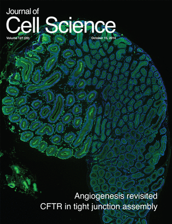
Immunofluorescent staining of CFTR (green) in the proximal mouse epididymis. In addition to being located in the apical membrane, CFTR is localised at tight junctions, where it uses its PDZ-binding domain to interact with ZO-1. Through this interaction, CFTR mediates the transcription of genes that control differentiation and proliferation, which it modulates by regulating the retention or recruitment of ZO-1 nucleic acid binding protein (ZONAB; also known as YBX3) to tight junctions. Blue, DAPI. See article by Ye Chun Ruan et al.

We undertook the present study to determine how epididymal phagocytes respond to the transient wave of apoptosis initiated by unilateral efferent duct ligation (EDL) in the epididymal epithelium. We show profound morphological and phenotypical changes restricted to the MPs populating the proximal epididymis following EDL.
E. R. Derbyshire, J. Clardy et al. explain how they completed this screen with a diverse array of kinase inhibitors, including several compounds already in clinical trials for cancer, to discover potential probes to interrogate the malaria kinome. Several different parasite kinases were identified as potential targets from analysis of the screening hits and the work demonstrates that the identified kinases are essential to both liver- and blood-parasite stages, thus highlighting the essential role of kinases to parasite biology. These findings identified small-molecule probes to explore malaria biology as well as show that the malaria kinome is a target rich for disease prevention and treatment.
In their Review Article, Ralph Weissleder and colleagues also discuss cancer imaging in relation to macrophage imaging — and extend their discussion into other disease areas that involve macrophage uptake and activity, namely, atherosclerosis, myocardial infarction, aortic aneurysm and diabetes
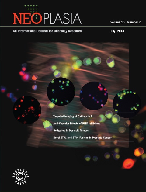
The cover is an artistic depiction of the results described in the paper by Keliher et al., wherein a nanomolar affinity small molecule targeting Cathepsin E allows for specific delineation of individual cancer cells allowing for more refined surgical staging, planning and resection with curative intent.

Pet MRI image of an apoE KO mouse. We also see the CT, which was used to fuse pet with MRI. The Pet signal reports on the nanoparticle uptake into plaque macrophages.

Modified radiotherapy fields based on vascular, rather than bony, anatomy were smaller than conventional fields in the treat- ment of seminoma and may allow for a significant reduction in the volume of normal tissue irradiated. This offers the potential of decreasing late effects in seminoma survivors without sacrificing disease coverage and cancer control. Validation of these results may inform a redefinition of the standard fields for radiotherapy and allow for more targeted and personalized radiation delivery in the treatment of seminoma.

The cover image is a digitally altered H&E staining of a mismatched (BALB/c into C57BL/6) heart allograft. The graft shown is from a C57BL/6 B7 double knockout recipient treated with an anti-B7-H3 mAb following transplantation. The result is more severe cellular rejection, with a dense chronic inflammatory infiltrate and extensive myocyte damage, as compared with control-treated mice. The image is taken from Ueno et al. in which B7-H3 signaling is shown to prolong graft survival; therefore, B7-H3 signaling appears to act as in a coinhibitory manner, reigning in the immune response. As B7-H3 signalling has in other systems been shown to be costimulatory, it seems that the outcome of B7-H3-signaling may be context dependent.
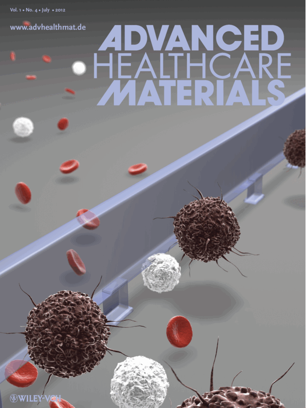
Many cancers shed malignant cells into the circulation. These cancer cells are important for both diagnostic and therapeutic purposes but their purification, quantification and characterization remain challenging. On page 432, Hakho Lee, Ralph Weissleder, and co-workers present a low-cost, rapid microfluidic cell sorter (μFCS) device for the detection and molecular analysis of circulating tumor cells (CTCs). The μFCS efficiently enriches CTCs from unprocessed whole blood, allows on-chip culture and molecular profiling, and provides cell retrieval for subsequent analyses. The potential clinical application of the technology is demonstrated by capturing and genetically analyzing CTCs in tumorbearing mice.

Flagging Down Tumor Cells. Cancer cells present in your bloodstream could indicate tumor metastasis to other sites in the body. To detect these rare, circulating tumor cells (CTCs), Issadore and colleagues attached magnetic nanoparticles to CTCs found in the blood of ovarian cancer patients. These cells were then flowed through a magnetic microdevice where they acquired a magnetic moment, as is colorfully depicted on this week's cover. The magnetic fields of the patients' CTCs were then identified and quantified. The hope is to eventually deploy this microdevice to the clinic, where CTC burden could correlate with cancer progression.
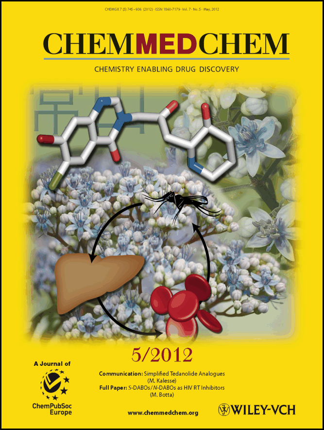
The cover picture shows a molecular model of the natural product analogue halofuginone and the life cycle of the malaria parasite that requires a mosquito vector, the sequential infection of mammalian liver cells, followed by the invasion of red blood cells. Halofuginone is a synthetic derivative of the plant alkaloid febrifugine, which constitutes the active component of “Chang Shan”—a traditional Chinese medicine prepared from the roots of the blue evergreen hydrangea Dichroa febrifuga. Both compounds have been widely recognized for their activity in the symptomatic blood stage of malaria. We have discovered that halofuginone also potently inhibits the early- and late-liver-stage processes, clearing parasites one day after liver cell infection, a feature that is uncommon for most antimalarial agents. This finding reveals that halofuginone has potential as a prophylactic that could prevent the progression of the disease to the life-threatening blood stage.
A number of small molecule PARP inhibitors are currently undergoing advanced clinical trials. Determining the distribution and target inhibitory activity of these drugs in individual subjects, however, has proven problematic. Here, we used a PARP agent for PET-CT imaging (18F-BO), which we developed based on the Olaparib scaffold using rapid bioorthogonal conjugation chemistries. Standard 18F-FDG imaging, however, failed to detect such therapy induced changes. This research represents a step towards developing a more generic approach for the rapid codevelopment of companion imaging agents based on small molecule therapeutic inhibitors.
Magnetic resonance imaging of magnetic nanoparticles allows monitoring of disease progression in type 1 diabetes. Mathis and colleagues (p 361; and News and Views by Chervonsky, p 311) use this approach to predict diabetes onset and identify a pathway important in the regulation of disease progression. The original image is a coronal view of an anesthetized mouse visualized by magnetic resonance imaging with a 4.7-Tesla microimaging system.

Go with the Flow. In healthy people, blood flows uninterrupted throughout the body. In patients with sickle cell disease, however, red blood cells take on a crescent shape that physically impedes the flow of blood. Wood et al. have created a microfluidic device that can measure such decreased flow velocity in blood samples from sickle cell patients. This biophysical indicator can then be used to stratify patients on the basis of disease severity and predict response to therapy.
Visualization of dendritic cells in the initial segment of a CD11c-EYFP mouse epididymis. Peritubular CD11c-positive cells project intraepithelial dendrites toward the lumen. Tight junctions are labeled with an anti-ZO-1 antibody (red), nuclei are shown in blue. Picture created using a Nikon ECLIPSE 90i microscope and NIS-Elements. Courtesy N Da Silva.

Current management of aortic aneurysms (AAs) relies primarily on size criteria to determine whether invasive repair is indicated to preempt rupture. We hypothesized that emerging molecular imaging tools could be used to more sensitively gauge local inflammation. Because macrophages are key effector cells that destabilize the extracellular matrix in the arterial wall, it seemed likely that they would represent suitable imaging targets. We here aimed to develop and validate macrophage-targeted nanoparticles labeled with fluorine-18 (18F) for positron emission tomography

Visualization of dendritic cells in the initial segment of a CD11c-EYFP mouse epididymis. Peritubular CD11c-positive cells project intraepithelial dendrites toward the lumen. Picture created using a Nikon ECLIPSE 90i microscope and NIS-Elements.

The figure shows a three-dimensional reconstruction of hybrid fluorescence molecular tomography/x-ray computed tomography (FMT-CT) in a mouse 3 days after coronary artery ligation. The red signal in the infarcted apical myocardium encodes the distribution of a nanoparticle targeted to monocyte/macrophages, which derivatized the fluorochrome. The multimodal imaging approach allows one to noninvasively quantitate infarct inflammation and the quality of tissue repair by optical tomography, while CT imaging provides the exact anatomic location of the molecular signal.

When Small Meets Smart. A miniaturized nuclear magnetic resonance (NMR) device detects scarce cancer cells in tissue biopsies with speed and accuracy, reported by Haun et al. in this issue. The coils generate radio frequency magnetic fields that excite protons in the tissue samples resulting in NMR signals that can be used to identify a cancer protein signature. This tiny portable micro-NMR machine operated by a smart phone can be used at the patient

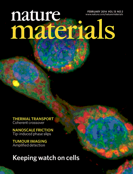
Imaging has become an indispensable tool in the study of cancer biology and in clinical prognosis and treatment. The rapid advances in high resolution fluorescent imaging at single cell level and MR/PET/CT image registration, combined with new molecular probes of cell types and metabolic states, will allow the physical scales imaged by each to be bridged. This holds the promise of translation of basic science insights at the single cell level to clinical application. In this article, we describe the recent advances in imaging at the macro- and micro-scale and how these advances are synergistic with new imaging agents, reporters, and labeling schemes. Examples of new insights derived from the different scales of imaging and relevant probes are discussed in the context of cancer progression and metastasis.

Chemical probes provide important tools for dynamically interrogating biological systems and for investigating potential drug targets. On the cover, we highlight three research papers in this issue that describe advances in chemical probe research: using new biochemical assays, Bradner et al. discover unexpected selectivity within current HDAC inhibitors and develop a true pan-HDAC inhibitor (p238); Kokel et al. describe a method for identifying neuroactive molecules and predicting their mode of action through behavioral zebrafish assays (p231); and Bracha et al. use a combination of RNAi, metabolomics and chemical probes to uncover metabolic enzymes that regulate myoblast differentiation (p202).

We implement the use of a graphics processing unit (GPU) in order to achieve real time data processing for high-throughput transmission optical projection tomography imaging. By implementing the GPU we have obtained a 300 fold performance enhancement in comparison to a CPU workstation implementation. This enables to obtain on-the-fly reconstructions enabling for high throughput imaging. Reconstructed image results for the absorption coefficient for 360 projections

The aim of this study was to iteratively develop and validate an 18F-labeled small vascular cell adhesion molecule (VCAM)-1 affinity ligand and demonstrate the feasibility of imaging VCAM-1 expression by positron emission tomography

Monocytes and macrophages play active roles in atherosclerosis, a chronic inflammatory disease that is a leading cause of death in the developed world. The prevailing paradigm states that, during human atherogenesis, monocytes accumulate in the arterial intima and differentiate into macrophages, which then ingest oxidized lipoproteins, secrete a diverse array of proinflammatory mediators, and eventually become foam cells, the key constituents of a vulnerable plaque. Yet monocytes are heterogeneous. In this review we summarize recent advances of our understanding of the behavioral heterogeneity of monocytes during disease progression and outline emerging molecular imaging approaches to address key questions in the field.

Collection of images highlighting the breadth of topics, techniques and model systems being showcased in this special issue on solute transport and acid−base regulation. Images courtesy of (clockwise from top left): Paul Linser; Winnie Shum; Jiun-Lin Horng, Li-Yih Lin and Pung-Pung Hwang; Daniel Hilger, Yevhen Polyhach, Etana Padan, Heinrich Jung and Gunnar Jeschke (reproduced with permission from Biophys. J. 93, 3675−3683); R. Todd Alexander and Sergio Grinstein; Winnie Shum; Dennis Brown, Teodor Paunescu, Sylvie Breton and Vladimir Marshansky; Max Schwanitz (centre image).
Tumor-associated macrophages (TAM) invade the tumor stroma in many cancers, yet their role is incompletely understood. To visualize and better understand these critical cells in tumor progression, we screened a portfolio of rationally selected, injectable agents to image endogenous TAM ubiquitously in three different cancer models (colon carcinoma, lung adenocarcinoma and soft tissue sarcoma). AMTA680, a functionally derivatized magnetofluorescent nanoparticle, labeled a subset of myeloid cells with a
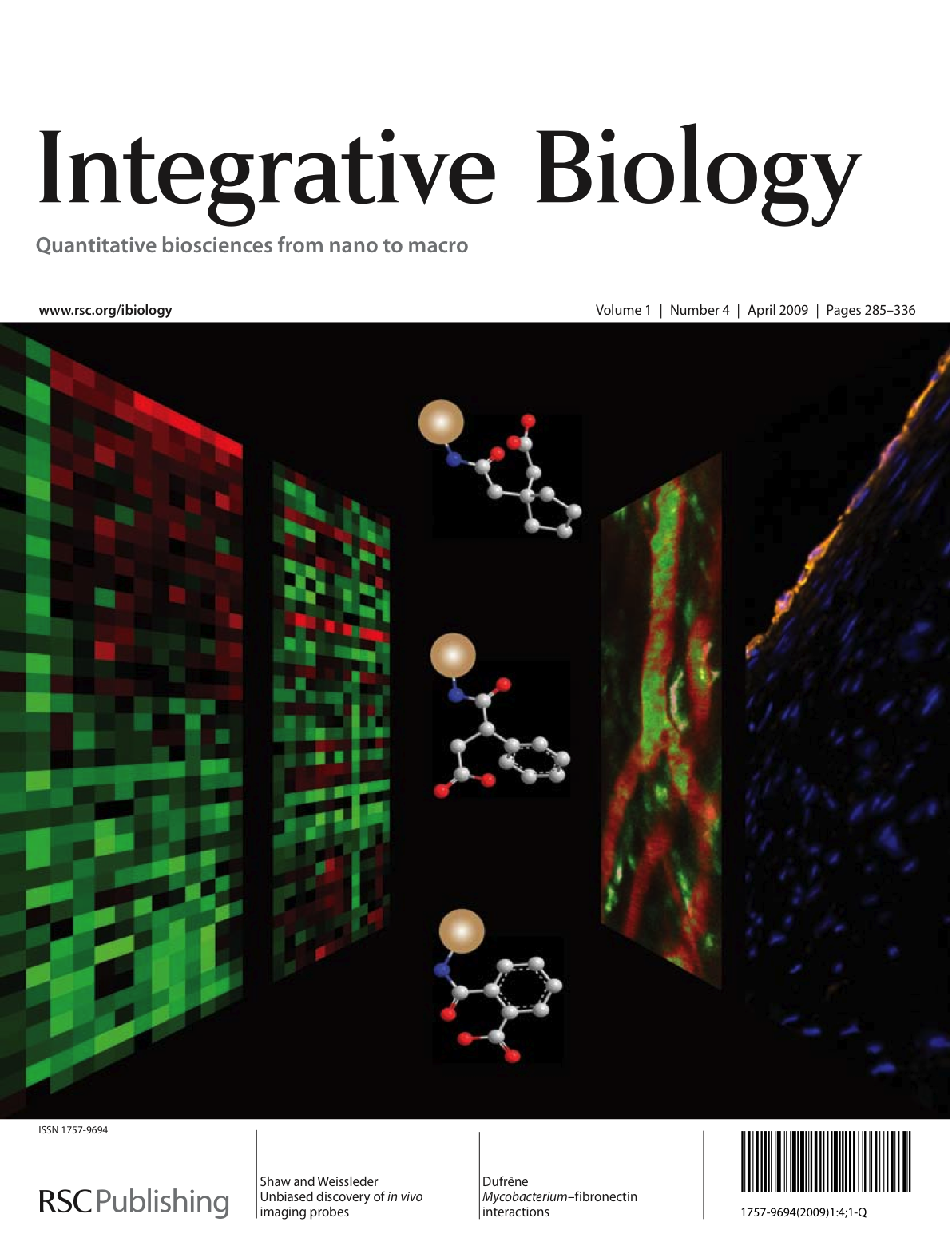
In vivo imaging reveals how proteins and cells function as part of complex regulatory networks in intact organisms, and thereby contributes to a systems-level understanding of biological processes. However, the development of novel in vivo imaging probes remains challenging. Here, we rapidly profile the in vitro binding of nanoparticle imaging probes in multiple samples of defined target vs. background cell types, using primary cell isolates. We apply this approach to the identification of nanoparticle imaging probes that bind endothelial cells, and validate our in vitro findings in human arterial samples, and by in vivo intravital microscopy in mice. Overall, this work presents a generalizable approach to the unbiased discovery of in vivo imaging probes, and may guide the further development of novel endothelial imaging probes

Special Issue: Nanoparticle enhanced imaging - Emerging oncologic applications.
Guest-Editor: Dr. Harisinghani

Osteoclasts degrade bone matrix by demineralization followed by degradation of type I collagen through secretion of the cysteine protease, cathepsin K. Current imaging modalities are insufficient for sensitive observation of osteoclast activity, and in vivo live imaging of osteoclast resorption of bone has yet to be demonstrated. Here, we describe a near-infrared fluorescence reporter probe whose activation by cathepsin K is shown in live osteoclast cells and in mouse models of development and osteoclast upregulation. Cathepsin K probe activity was monitored in live osteoclast cultures and correlates with cathepsin K gene expression. In ovariectomized mice, cathepsin K probe upregulation precedes detection of bone loss by micro-computed tomography. These results are the first to demonstrate non-invasive visualization of bone degrading enzymes in models of accelerated bone loss, and may provide a means for early diagnosis of upregulated resorption and rapid feedback on efficacy of treatment protocols prior to significant loss of bone in the patient.
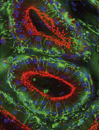
From �Segmental Expression of the Bradykinin Type 2 Receptor in Rat Efferent Ducts and Epididymis and Its Role in the Regulation of Aquaporin 9� by C. Belleann�e, N. Da Silva, W. W. C. Shum, M. Marsolais, R. Laprade, D. Brown, and S. Breton. Localization of the bradykinin type 2 receptor (BDKRB2) in rat cauda epididymidis and efferent ducts. BDKRB2 (red) is localized in the apical membrane of epididymal principal cells. Clear cells, which express the E subunit of the proton pumping V-ATPase (green), are negative for BDKRB2. In the smaller efferent duct tubules shown in the bottom left corner, BDKRB2 is localized in the apical membrane of non-ciliated cells (yellow) and is absent from ciliated cells, identified by their positive labeling for the anion exchanger AE2 in their basolateral membrane (green). Nuclei are visualized in blue with DAPI. Belleann�e et al. show that luminal bradykinin activates the aquaglyceroporin, AQP9, in the distal initial segment of the epididymis. Cover design and layout: Jane Tenenbaum, Tenenbaum Design, Cambridge, Massachusetts. Biology of Reproduction is printed with soy-based inks on acid-free paper by Allen Press, Inc., Lawrence, Kansas.

New technologies for imaging molecules, particularly optical technologies, are increasingly being used to understand the complexity, diversity and in vivo behaviour of cancers. Omic approaches are providing comprehensive snapshots of biological indicators, or biomarkers, of cancer, but imaging can take this information a step further, showing the activity of these markers in vivo and how their location changes over time. Advances in experimental and clinical imaging are likely to improve how cancer is understood at a systems level and, ultimately, should enable doctors not only to locate tumours but also to assess the activity of the biological processes within these tumours and to provide on the spot treatment.

The existing paradigms for how immune cells interact with each other and their environment have come largely from static images obtained through histochemistry. As this volume illustrates, an exciting era in immunology is now emerging where the immune response and much of its complexity can be visualized in real time and within the confines of secondary lymphoid tissues in vivo.
As the reviews in this issue describe, such studies are altering our perceptions of how, when, where, and for how long immune cells participate in innate and adaptive responses. Our authors show how the advances in and applications of new technologies facilitating imaging of the immune system are destroying old paradigms and writing new ones.

In this study we report on the use of a new, bolus injectable, carboxymethyl-dextran based magnetic nanoparticle (MNP), ferumoxytol, to improve detection in loco-regional lymph nodes by magnetic resonance imaging (MRI). Conclusion: Ferumoxytol is safe and, at the appropriate circulation interval, modulates nodal signal intensity, allowing for identification of malignant nodal involvement by MRI.
Double-immunostaining of V-ATPase B1 knockout mouse epididymis showing aquaporin 9 (green) and V-ATPase B2 (red). Nuclei are stained with DAPI (blue). From: Da Silva N, Shum WWC., El-AnnanJ, Paunescu TG, McKee M, Smith PJS, Brown D, and Breton S. "Relocalization of the V-ATPase B2 subunit to the apical membrane of epididymal clear cells of mice deficient in the B1 subunit."

Restoring endogenous p53 expression leads to regression of autochthonous lymphomas and sarcomas in mice without affecting normal tissues. The mechanism responsible for tumour regression is dependent on the tumour type, with the main consequence of p53 restoration being apoptosis in lymphomas and suppression of cell growth with features of cellular senescence in sarcomas. These results support efforts to treat human cancers by way of pharmacological reactivation of p53.

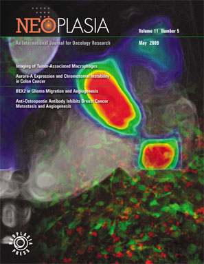
Colored scanning EM of a human macrophage, the key player in 3 articles featured in the current issue. Lumeng et al. describe a novel subset of macrophages resident in adipose tissue (page 175). Tacke and colleagues (page 185) and Swirski et al. (page 195) both delve further into the roles macrophages play in atherosclerotic plaques. Siamon Gordon synthesizes the findings of the 3 papers in a Commentary beginning on page 89.

This study thus provides a foundation for using Magnetofluorescent nanoparticles (MFNPs) to image genetic and/or pharmacological perturbations of cellular inflammation in experimental atherosclerosis and for the future development of novel targeted nanomaterials for atherosclerosis.

Molecular and cellular mechanisms of atherogenesis and its treatment are largely being unraveled by in vitro techniques. We describe methodology to directly image macrophage cell activity in vivo in a murine model of atherosclerosis using laser scanning fluorescence microscopy (LSFM) and a macrophage-targeted, near-infrared fluorescent (NIRF) magnetofluorescent nanoparticle (MFNP)

A novel magnetic resonance lymphangiographic technique was employed to highlight the likely sites of occult nodal metastasis from prostate cancer. Conclusions: Nodal metastases from prostate cancer are largely localized along the major pelvic vasculature. Defining nodal radiation treatment portals based on vascular rather than bony anatomy may allow for a significant decrease in normal pelvic tissue irradiation and its associated toxicities.

This study was designed to investigate the role of activated H-RAS on the angiogenic phenotype of melanoma that arises in the inducible Tyr/Tet-RAS Ink4a/Arf(-/-) model using in vivo imaging with histopathologic correlation. We show that loss of RAS activity in fully established melanomas led to a reduction in tumor volume, which was preceded by impairment of vascular function as determined by in vivo magnetic resonance imaging.

Here we describe the properties of a luciferase from the copepod marine organism Gaussia princeps. Gaussia luciferase provides a sensitive means of imaging gene delivery and other events in living cells in culture and in vivo, with a unique combination of features including high signal intensity, secretion, and ATP independence, thus being able to report from the cells and their environment in real time.
We have developed an imaging agent for real-time endoscopic tumor detection in a murine model using a previously identified phage library-derived colon cancer-specific cyclic peptide and fluorescent moieties. The modified peptide had a 24 minute blood half life and tumoral accumulation was 6.9% of injected dose/g, 7-fold higher than a scrambled control peptide. These results show proof-of-principle that disease-specific library-derived fluorescent probes can be rapidly developed for use in the early detection of cancers by optical means.
Recent studies have described neuronal progenitor cell recruitment to tumors in vivo, however, the mechanisms mediating this recruitment are not yet understood. Our data suggest that recruitment of C17.2-luc cells to TEC is mediated via SDF- 1a/CXCR4 activation that results in modification of a4-integrin and results in improved recruitment of C17.2-luc cells.

Here we report on the synthesis of a novel imaging probe that is specific for HIV-1 protease (PR). The probe was designed to be biocompatible, i.v. injectable, and detectable by fluorescence imaging. These results are the first proof of principle that viral proteases can directly be imaged in vivo.

Using a model of gliosarcoma with stably green fluorescence protein-expressing 9L glioma cells, we explored a multimodal (near-infrared fluorescent and magnetic) nanoparticle as a preoperative magnetic resonance imaging contrast agent and intraoperative optical probe. This prototypical multimodal nanoparticle has unique properties that may allow radiologists and neurosurgeons to see the same probe in the same cells and may offer a new approach for obtaining tumor margins.

We developed a novel, biocompatible, and physiologically inert nanoparticle (highly derivatized cross-linked iron oxide nanoparticle; CLIO-HD) for highly efficient intracellular labeling of a variety of cell types that now allows in vivo MRI tracking of systemically injected cells at near single-cell resolution. Using B16-OVA melanoma and CLIO-HD-labeled OVA-specific CD8+ T cells, we have demonstrated for the first time high resolution imaging of T-cell recruitment to intact tumors in vivo.

The paper by Ntziachristos, et al. describes the use of fluorescence-mediated tomography (FMT) imaging of deep tissues to generate high resolution, quantitative images of biochemical events in living animals. The capability of FMT to provide 3-dimensional images of biochemical events should play a significant role in the development and use of optical imaging technologies in biomedicine.

Contrast enhanced, near infrared optical tomography has the potential to provide a wealth of structural and functional information about biological tissue. The cover artistically illustrates contrast-enhanced NIR tomography when taking measurements with an intensity-modulated source. The photon density wavefronts propagate from the source fiber optic to an obscured, elliptic inclusion within the tissue. Exhibiting enhanced contrast via a specialized fluorescent contrast agent, a series of fluorescent photon density wavefronts propagate from the inclusion to a fiber optic detector, which receives the diagnostic information.