Positron emission tomography – computed tomography (PET-CT) imaging facility
The primary use for the PET-CT system is in the development and validation of novel radiolabeled positron-emitting ligands, which can be used for tumor detection, diagnosis of cardiovascular and autoimmune disease or for establishing metabolic pathways.
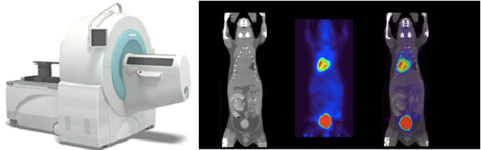
Siemens Inveon PET-CT
The new Siemens Inveon PET-CT (formerly Concord Focus) system uses high light output LSO (Lutetium Oxyorthosilicate) crystals, and has a timing resolution of less than 1.5 nsec, a greater than 10% peak absolute sensitivity, a stationary field of view (FOV) of 12.7 cm (which can increase to 30 cm FOV with continuous bed motion), an energy resolution of less than 18%, over 25,000 individual detector elements, and a spatial resolution of less than 1.4 mm. The CT uses CCD technology that allows the highest available signal-to-noise ratio, and fiber optics that permit the highest efficiency light collection. It has 4,064 × 4,064 detectors, a FOV greater than 10 × 10 cm, a spatial resolution of 15 micron isotropic voxels, and can scan an entire mouse in less than 1 minute.
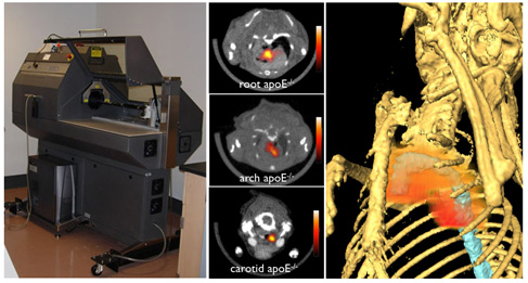
GammaMedica X-PET
The imaging system used is the Gamma Medica FLEX™ small animal imaging system with X-PET™ and X-O™ modalities. The X-PET system is a high sensitivity / large axial field of view small animal PET scanner. The three-dimensional PET system is comprised of 11, 520 (2.3 mm x 2.38 mm x 10 mm) Bismuth Germanate (BGO) crystals in 48 separate rings. The large axial field of view (11.6 cm) can image an entire mouse with a resolution of less than 2 mm.