CT imaging facility
Computed tomography is used in a variety of ways at the CSB (MGH). It is often used as a stand-alone device for tumor and lesion detection, angiography, and for making diagnoses e.g. for myocardial infarction or bone degeneration. It is also used in combination with other imaging modalities such as Positron Emission Tomography (PET), Single Photon Emission Computed Tomography (SPECT), and Fluorescence Molecular Tomography (FMT) to provide an anatomical framework with which to register the functional data.
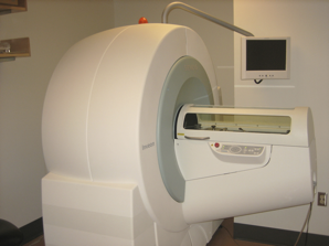
Siemens Inveon PET-CT
The primary CT system is part of the Siemens Inveon PET-CT, which utilizes CCD technology and allows the highest available signal-to-noise ratio. It also makes use of fiber optics, which permit the highest efficiency light collection. The Inveon system has 2,048 × 3,072 detectors, a field of view of up to 11 × 8.5 cm, a spatial resolution of 40 micron isotropic voxels, and can scan an entire mouse inless than 5 minutes.
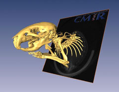
The Inveon has been used to characterize bone structure (such as examining osteolysis in cancer), and has also quantified mineral densities in transgenic mouse models. In addition, the Inveon frequently provides anatomical CT reference images for other modalities, such as for bi-modal FMT-CT data sets or three-dimensional virtual histology.
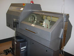
GammaMedica X-SPECT
A second CT system is housed within the X-SPECT / X-O system. It is a 50 kVp, 1.0 mA, fixed anode and air cooled X-ray tube with a 2-inch x 2-inch (48 µm pitch) solid state detector (1024 × 1024 array).
GammaMedica X-PET
The third CT system is contained within the XPET / XO system. It has a 80 kVp, 0.6 mA, fixed anode and air cooled X-ray tube with a 4.72-inch x 4.72-inch (50 µm pitch) solid state dectector (2048 × 2048 array). This later system is capable of imaging an entire mouse in less than a minute.