In the course of treating cancer, oncologists are keenly interested in understanding the molecular features of their patients’ tumors: to know what specific proteins are present, which growth-promoting pathways are active, and therefore what the optimal treatment(s) for each patient might be.
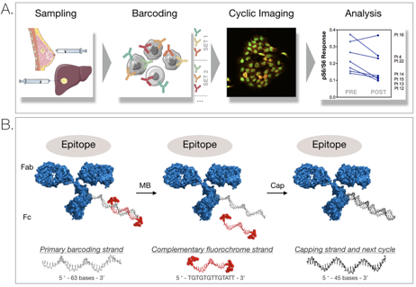
One common method of obtaining samples from patients tumors is called a fine needle aspirate (Figure 1A), where a thin needle is inserted into the biopsy site and tumor cells are removed for analysis. This method has several advantages: firstly, due to the small size of the needle it subjects the patient to less pain and less medical risk, secondly, it can more easily be performed in an outpatient setting and thirdly, results from the biopsy can typically be more rapidly obtained.
In an effort to further expand the usefulness of fine needle aspirates beyond single protein assays, CSB scientists have developed an approach called “SCANT”, which uses antibody-DNA conjugates to efficiently “cycle” through the detection of multiple proteins of interest, dramatically expanding the number of targets that can be analyzed from a single fine needle aspirate (Figure 1). With this method, it is possible to obtain both more information from patient samples with relatively few cells, as well as to probe cancer pathways in far greater depth than currently occurs for routine biopsy samples in the clinic.
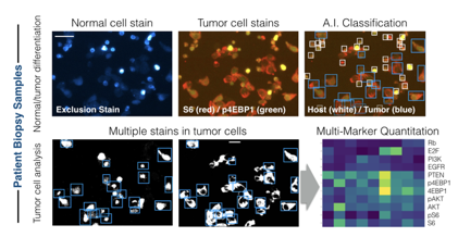
Figure 2: Single Cell Analysis Workflow for patient biopsy samples. Tumor cells from the biopsy are gently separated into single cell clusters and then transferred onto glass slides and prepared for SCANT cyclic imaging. Following staining, a neural-network artificial intelligence (A.I.) algorithm identifies both host cells and tumor cells, including their boundaries, allowing measurement of the key target proteins across multiple cycles of imaging.
Utilizing this technology, the team from CSB analyzed the PI3-Kinase (PI3K) pathway (a growth-promoting signal commonly found to be important in many cancers) in patients who were participating in clinical trial of a new breast cancer combination therapy that included a PI3K-inhibitor. Using the SCANT method, preliminary results in a small group of patients fine needle aspirates showed that many of the key signaling proteins expected to be altered from treatment indeed had measurable differences detected by SCANT. These preliminary results provide a rationale for further testing of the method in clinical trials and with potentially other pathways. These tools offer the hope of transforming fine needle aspirates into a key tool for guiding the selection and measuring the response of patients to targeted treatments in cancer therapy.
Article
Giedt RJ, Pathania D, Carlson JCT, McFarland PJ, Del Castillo AF, Juric D, Weissleder R
Single-cell barcode analysis provides a rapid readout of cellular signaling pathways in clinical specimens
Nat Commun. 2018;9(1):4550 – PMID: 30382095 – PMCID: PMC6208406 – DOI: 10.1038/s41467-018-07002-6