The Pathology Core is located on the 5th floor of the Simches Research Building (room 5178) and has laboratory space assigned for sectioning, staining and mounting.
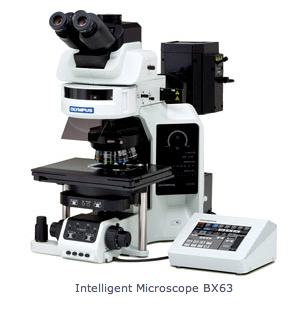
Olympus BX63
The upright automated Olympus BX63 microscope is equipped with a color CCD camera and MetaMorph software for imagining of histology preparations. It is equipped with 4, 10, 20, 40, 60 (OIL), 100 (OIL)x objectives, and the following filter cubes:
- DAPI-1160B-OFF-ZERO: EX 387/11, DM 409, EM 447/60
- GFP-3035D-OFF-ZERO: EX 472/30, DM 495, EM 520/35
- TXRED-4040C-OFF-ZERO: EX 562/40, DM 593, EM 624/40
- CY3-4040C-OFF-ZERO: EX 531/40, DM 562, EM 593/40
- CY5-4040B-OFF-ZERO: EX 628/40, DM 660, EM 692/40

Nikon Eclipse E400
The upright Nikon Eclipse E400 is equipped with a color CCD camera (Insight) and Spot software, as well as with 4, 10, 40, 60 and 100x objectives for still digital imaging of stained histology preparations.

Axiovert 100TV
The inverted Axiovert 100TV Zeiss microscope is equipped for fluorescence with filter cubes for FITC/GFP (fluorescein isothiocyanate/green fluorescent protein), Rhodamine/Cy3 and Cy5.5. It also has 10, 20 (phase), 40 (phase), 60 and 100x objectives. The microscope is equipped with a low-light video rate camera for live time digital imaging under transillumination/phase contrast and a Photometrics CoolSnap HQ CCD camera for still digital fluorescence images, both are coupled to a G4 Macintosh computer running IP Labs software and ImageJ.

Nikon 80i
The upright Nikon 80i epifluorescence microscope is equipped with 10x (phase), 20x, 40x and 60x Plan Fluor short working distance objectives, and the following filter cubes:
- #96300 UV-1A DAPI filter set: Excitation 360-370, Dichroic Mirror DM380, Emission LP420
- #96320 FITC/GFP HyQ filter set: Excitation 460-500, Dichroic Mirror DM505, Emission 510-560
- #96305 G-1B filter set: Excitation 541-551, Dichroic Mirror DM565, Emission LP590
- #41023 Cy5.5 filter set: Excitation 627-672, Dichroic Mirror DM685, Emission 685-735
- #41009 Cy7/AF750 filter set: Excitation 673-748, Dichroic Mirror DM750, Emission 765-835
It is equipped with a Photometrics Cascade low intensity back-illuminated CCD camera coupled to a G4 Macintosh computer running IP Labs software. The microscope also has live focus capability under both transillumination and fluorescence with a 100 millisecond refresh rate.

Nikon TE2000
The inverted Nikon TE2000 epifluorescence microscope is equipped with 4, 10, 20 and 40x phase/fluorescence objectives as well as with GFP/FITC, Rhodamine/Cy3, Cy5, Cy5.5 and Cy7 filter cubes. It is also equipped with a color Insight CCD camera and Spot Software for digital imaging of live cells.

Nikon Eclipse 50i
- The upright Nikon Eclipse 50i Light/Fluorescence is equipped with a color CCD camera (Insight) and Spot software (4.1) 4, 10, 20, 40 and 100x objectives for still digital imaging of stained histology preparations.
- The upright Nikon Eclipse 50i Light microscope is equipped with 4, 10, 20, 40 and 100x objectives for imaging of stained histology preparations.

Leica (CM1900) Rapid Sectioning Cryostat
- Incorporates two separate cooling systems; features specimen cooling adjustable to -50 degrees C and cabinet cooling to -35 degrees C.
- Section thickness can be set between 1 to 60μm.
- A 90° prism head mounts onto the specimen head for quick freezing of specimens in place.

Services
Histology services: bold indicates routine work as services
- Dissecting, harvesting, embedding of tissues
- Sectioning (frozen)
- Tissue banking
- Quantification analysis
- Hematoxylin and eosin (H&E) staining
- Trichrome staining (MASSON)
- Immunohistochemistry
- Immunofluorescent staining
- Alkaline Phosphatase staining (ALP)
- Von Kossa staining (VK)
- Gram staining
- Elastic staining
- Oil Red O staining
- Picrosirius Red Polarization
- Prussian Blue staining
- Periodic Acid-Schiff staining
- Acid Phosphatase Leukocyte (TRAP) staining
- Enzyme-linked immunosorbent assay (ELISA)
- Reverse transcription-polymerase chain reaction (RT-PCR)
- In situ hybridization (ISH)
- Fluorescent in situ hybridization (FISH)
- Tissue microarrays
- Western blotting
- Sectioning (paraffin)